
Prostate Cancer
Diagnosis / Screening
Regular screening facilitates early intervention - and early cures
Understanding Prostate Cancer
Many men, even from their 30s onwards, can have very small, slow-growing prostate cancers that never cause harm. Autopsy studies show that these tiny cancers become more common with age, affecting a large proportion of men by their 60s and beyond. The challenge is to identify significant cancers that could affect your health, while avoiding unnecessary anxiety and treatment for harmless ones.
What counts as “significant” depends on your age and overall health. A prostate cancer that warrants treatment in a fit 60-year-old may be less important for someone in their 80s with other medical conditions.
PSA/ Prostate Specific Antigen
Prostate-specific antigen (PSA) is a protein produced by the prostate. Most is released into semen, but small amounts enter the bloodstream, where it can be measured with a simple blood test. PSA itself is not harmful and does not cause cancer; it is one piece of information used to estimate the likelihood of prostate cancer.
An elevated PSA does not necessarily indicate cancer. Levels may rise due to prostate enlargement, inflammation, infection, or increasing age. Conversely, a normal PSA does not exclude cancer. Higher values are generally more concerning, although what constitutes a “raised” PSA varies with age, family history, and other risk factors—often around 3.
PSA is only part of the overall assessment. Interpretation also considers family history, DRE findings, PSA trends over time, prostate size, and imaging such as MRI.
Bottom line
PSA is a useful first step, but not the whole picture. It is interpreted alongside clinical history, examination, and imaging to guide appropriate next steps.
Download our patient fact sheet: Prostate Specific Antigen (PSA)
Digital Rectal Examination (DRE)
The prostate sits just below the bladder, surrounds the urethra, and lies in front of the rectum. During a digital rectal examination (DRE), your doctor inserts a gloved finger into the rectum to assess the prostate. Most early prostate cancers cannot be felt, but a firm or irregular area may raise concern for malignancy.
If the examination is suspicious, or if your PSA is elevated, an MRI is usually the next step.
Prostate MRI
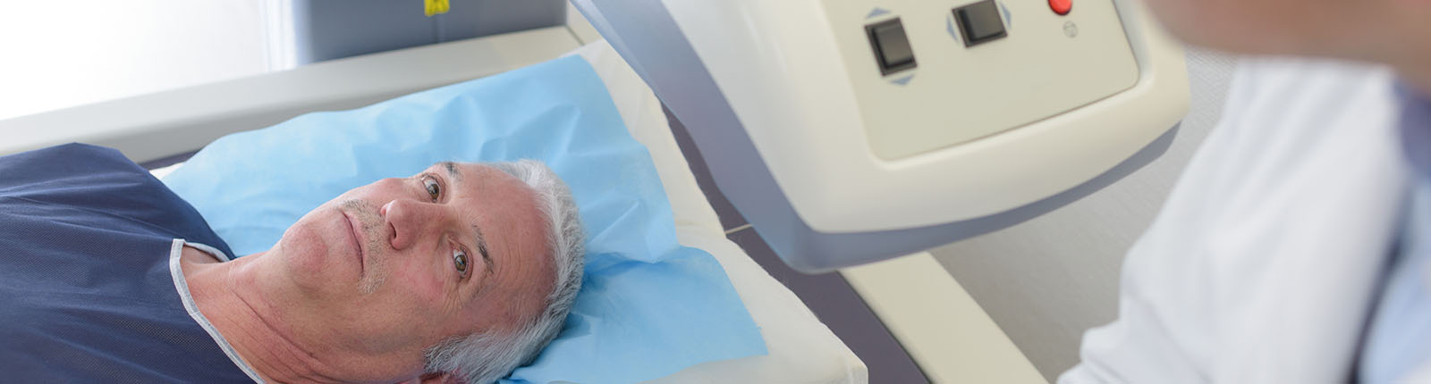
Multi-parametric MRI (mpMRI) is a non-invasive scan that uses a powerful magnetic field—not radiation—to produce detailed images of the prostate. It does not diagnose cancer on its own, but helps determine whether a biopsy is needed by identifying areas that appear suspicious.
The technique was validated in Australia at the Wesley Hospital in Brisbane in collaboration with Professor Jelle Barentsz (Radboud University, Nijmegen, Netherlands), a world leader in mpMRI. mpMRI-informed biopsy has since become standard practice worldwide. iMed Buderim, working in conjunction with iMed Wesley—one of the world’s most experienced mpMRI providers—is my preferred provider on the Sunshine Coast.
What to expect
You will lie still on the MRI table for approximately 30–40 minutes. Results are reported using a standard scoring system (PI-RADS 1–5), which reflects how suspicious an area appears. We will review the results together at a follow-up appointment.
Understanding your result
MRI results are not simply “clear” or “cancer”—they require interpretation. Suspicious areas are not always cancer, and some cancers may not be visible on MRI.
If a suspicious area is identified, a targeted biopsy can be arranged. This combines MRI images with real-time ultrasound in theatre, using robotic guidance to improve accuracy.
If the scan is clear, monitoring your PSA may be sufficient. However, if other risk factors—such as family history, DRE findings, or PSA density—raise concern, further evaluation is sometimes warranted. In this setting, a PSMA PET scan is usually the next step. Biopsy would then be considered only if the PSMA scan shows an abnormality, or, rarely, if clinical suspicion remains high despite both scans being normal.
Why have an MRI if a biopsy may still be needed?
Even when a biopsy is recommended regardless, MRI plays a critical role. A positive MRI helps guide precisely where biopsies should be taken, reducing the risk of missing significant cancer. It also provides valuable information about tumour grade and extent, which can influence treatment decisions.
Robotic Assisted Transperineal Prostate Biopsies
A biopsy involves taking small tissue samples for examination under a microscope and is the only way to confirm a diagnosis of early prostate cancer. It is typically recommended when PSA is elevated, the DRE is abnormal, or MRI identifies a suspicious area.
The transperineal approach—where needles pass through the skin between the scrotum and anus—has largely replaced the older transrectal method, reducing the risk of infection to around 0.2%. The procedure is performed under general anaesthetic and uses MRI–ultrasound fusion with robotic guidance to maximise accuracy.
If you've been booked in for a biopsy, see Preparing for Biopsy for what to expect, how to prepare, and what happens afterwards.
Staging and Grading
Treatment options and recommendations are guided primarily by the stage and grade of the cancer.
Stage describes how far the cancer has spread within or beyond the prostate:
-
Stage T1: The tumour appears confined to the prostate, and the gland feels normal on examination.
-
Stage T2: The tumour appears confined to the prostate, but the gland feels abnormal on examination.
-
Stage T3: The tumour has extended beyond the prostate capsule into nearby tissue and/or the seminal vesicles.
-
Stage T4: The tumour has invaded adjacent organs such as the bladder, rectum, or pelvic side wall.
Prostate cancer can also spread to lymph nodes and distant sites, particularly bone and other organs.
Early prostate cancer typically causes no symptoms. When advanced, symptoms may include fatigue, weight loss, bone pain, and urinary difficulties. For this reason, PSA testing plays a key role in detecting prostate cancer before it spreads.
Grade describes how aggressive the cancer appears under the microscope. Prostate cancer is now classified using the Grade Group system (1–5), based on the Gleason score.
Grade Group 1: Gleason
Grade Group 2: Gleason
Grade Group 3: Gleason
Grade Group 4: Gleason 4+4=8
Grade Group 5: Gleason , , or
This system replaces the older Gleason scale out of 10, where the lowest practical score was 6, which many patients found confusing.
In general, Grade Group 1 represents low-risk disease and is often suitable for active surveillance. Grade Groups 2 and 3 are intermediate risk, where treatment is often, but not always, recommended. Grade Groups 4 and 5 are high risk and typically more aggressive.
Tests Involved in Prostate Cancer Treatment Decisions
Gallium-68 PSMA scan
This study is performed in two parts: a CT scan followed by the PSMA PET scan. The CT uses a rotating X-ray and iodine-based contrast to create cross-sectional images. The contrast may cause a brief warm sensation; inform your doctor if you have a contrast allergy or asthma.
The PSMA component uses a radioactive tracer that binds to prostate cancer cells, combined with PET imaging to detect areas of disease.
PSMA is primarily used to assess whether cancer has spread beyond the prostate. In selected cases, it may also be used before biopsy—particularly if MRI is normal but other factors suggest moderate to high risk—to help determine whether biopsy is warranted. It is more sensitive than CT or bone scan alone, although approximately 5% of cancers may not be PSMA-avid.
No fasting is required, but good hydration is recommended. As the scan involves two stages with a delay to allow tracer distribution, the total appointment time is approximately 2.5 hours. It is not currently Medicare-funded and is generally reserved for higher-risk cases or when results are likely to influence management.
Bone scan
A bone scan may be used to assess whether prostate cancer has spread to the skeleton.
A small amount of radioactive tracer is injected into a vein. Areas of abnormal bone activity absorb more tracer and appear as “hot spots” on imaging. Scanning is typically performed about one hour after injection.
The level of radioactivity is low and considered safe. However, bone scans are not specific for cancer and may also detect other conditions such as arthritis. Additional imaging (e.g. X-ray or MRI) may be required to clarify abnormal findings.